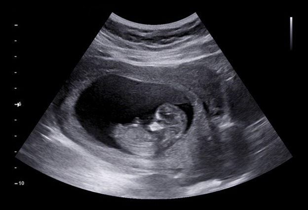
Pregnancy ultrasound allows healthcare experts to detect existing issues in the developing fetus and will enable parents to see their baby for the first time. As a result, ultrasounds are now an integral part of most pregnant women’s routine prenatal care.
According to The American Congress of Obstetricians and Gynecologists, diagnostic ultrasound methods are deemed safe and have no negative consequences on the growing fetus. However, ultrasounds should only be performed under the direction of an expert clinician.
Read on to learn about prenatal ultrasounds’ benefits, types, and significance and when they should be performed.
What exactly is a pregnancy ultrasound?
A pregnancy ultrasound is a test that images the developing baby and the mother’s reproductive organs using high-frequency sound waves. With each pregnancy, the typical number of ultrasounds varies.
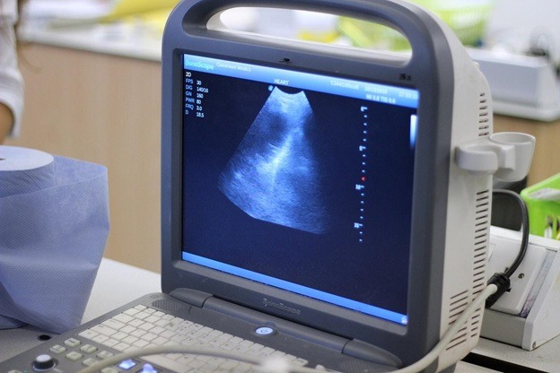
An ultrasound, often known as a sonogram, can help monitor normal fetal development and screen for potential issues. Along with routine ultrasound, there are a variety of more advanced ultrasounds, such as 3-D ultrasound, 4-D ultrasound, and fetal echocardiogram, which is an ultrasound that examines the fetus’ heart in depth. But a doctor or physician must look for the best ultrasound machine for sale that provides detailed images of the fetus’ heart.
Pregnancy ultrasound: Reasons
During pregnancy, an ultrasound can be utilised for a variety of purposes. If a previous ultrasound or blood test revealed a concern, your doctor might schedule more ultrasounds. Nonmedical ultrasounds may also be performed, such as to provide images for the parents or to determine the sex of the baby. While ultrasound technology is safe for both mother and child, healthcare providers advise against using it when there is no medical purpose or advantage.
During pregnancy’s first trimester
Ultrasounds may be performed during the first trimester of pregnancy (weeks one to twelve) to:
- pregnancy confirmation
- Examine the fetal heartbeat
- establish the baby’s gestational age and an estimated due date
- Examine for multiple pregnancies
- Examine the placenta, the uterus, the ovaries, and the cervix.
- determine whether the pregnancy is an ectopic pregnancy (when the fetus does not adhere to the uterus) or a miscarriage
- Examine the fetus for any abnormal growth.
During pregnancy’s second and third trimesters
An ultrasound may be performed throughout the second trimester (12 to 24 weeks) and the third trimester (24 to 40 weeks or birth) to:
- keep track of the fetus’s growth and position (breech, transverse, cephalic, or optimal)
- establish the gender of the baby
- confirm several pregnancies
- Examine the placenta for issues including placenta previa (when the placenta blocks the cervix) and placental abruption (when the placenta separates from the uterus before delivery)
- Look for Down syndrome traits (done between 13 and 14 weeks)
- Examine for congenital anomalies or birth problems.
- Look for structural abnormalities or blood flow issues in the fetus.
- monitor the amniotic fluid levels
- evaluate whether the fetus is receiving enough oxygen
- detect ovaries or uterine problems, such as pregnancy tumours
- determine the length of the cervix
- assist in the development of other tests, such as amniocentesis
- establish an intrauterine death

How to Get Ready for an Ultrasound
Earlier in the pregnancy, an ultrasound may require you to have a full bladder for the technician to gain a clear view of the fetus and your reproductive organs. One hour before your ultrasound, consume two to three eight-ounce glasses of water. It would help if you did not urinate before your ultrasound to arrive with a full bladder.
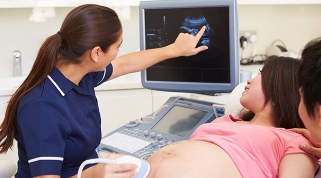
What occurs during an ultrasound?
You lie down on an examining table or bed during an ultrasound. An ultrasound technologist applies a special gel to your abdominal and pelvic area. Because the gel is water-based, it should not leave stains on your clothes or skin. The gel aids in the proper propagation of sound waves. The technician will next place a little wand called a transducer on your tummy. They move the transducer to capture images in black and white on the ultrasound screen. The technician may also take measurements of the screen image. They may request that you move or hold your breath while they take photographs. The technician then verifies that the relevant photos were collected and are transparent. The technician then wipes away the gel, allowing you to empty your bladder.
Pregnancy ultrasound types
More advanced ultrasound techniques may be used when a more detailed image is necessary. If the doctor detects abnormalities during your standard ultrasound, these may provide the information needed to make a diagnosis.
Ultrasound transvaginal
To get a sharper view, a transvaginal ultrasound may be performed. This ultrasound is more likely to be utilised early in pregnancy, when producing a clear image may be more difficult. A tiny ultrasound probe is placed into the vagina for this exam. While the photos are being gained, the probe is resting on the rear of your vagina.
3-D ultrasound
A 3-D ultrasound, unlike a typical 2-D ultrasound, allows your doctor to see the width, height, and depth of the fetus and your organs. This ultrasound can be extremely useful in diagnosing any abnormalities that may arise throughout your pregnancy. A 3-D ultrasound employs the same method as a regular ultrasound but creates the 3-D image with a special probe and software. It also involves specialised technician training. Thus, it may not be as commonly available.
4-D ultrasound
A dynamic 3-D ultrasound is another name for a 4-D ultrasound. It improves the image of the baby’s face and movements. This ultrasound is performed similarly to regular ultrasounds but using specialised equipment.
Echocardiography of the fetus
A fetal echocardiogram is performed if your doctor suspects your baby has congenital cardiac abnormalities. This test may be performed as a regular prenatal ultrasound. However, it may take longer to complete. It captures an in-depth image of the fetus’ heart, revealing its size, shape, and structure. This ultrasound also allows your doctor to see how your baby’s heart works, aiding in diagnosing heart abnormalities.
Final Words
Ultrasound is an ever-grown technology. Like any other test, the results might not be 100% accurate. However, the HMI x-ray can give parents and healthcare providers valuable information and services to help them manage and care for their baby and the pregnancy with the best ultrasound technology. Ultrasound allows parents to see their babies before they give birth. That helps them bond and establish a relationship.